


2. 中国科学院 理化技术研究所, 北京 100190
2. Technical Institute of Physics and Chemistry, Chinese Academy of Sciences, Beijing 100190, P. R. China
硫氧化钆(Gd2O2S)是一种重要的稀土硫氧化物,具有六方晶系结构和较宽的禁带宽度(4.2~4.8 eV),作为发光材料基质有非常高的光吸收和传能效率,掺杂激活离子后有很高的发光效率,可广泛应用于白光LED、医学影像、X射线成像、上转换发光和激光器等领域[1-6]。
近年来,文献报道了多种合成微纳米荧光粉的方法,主要包括:高温固相法[7-9]、共沉淀法[10, 11]、燃烧法[12, 13]、微波辅助法[14, 15]、微乳液法[16, 17]、热分解法[18]和水热/溶剂热法[19-21]等。上述方法各有优劣,如:共沉淀法主要优点在于制备工艺简单、成本低、条件易于控制、合成周期短和组成均匀等,缺点也很明显,即加入沉淀剂可能会使反应体系局部溶液浓度过高,导致颗粒产生团聚而造成产物组成不均一,且易于引入杂质而难以制备粒径较小的纳米颗粒。微波辅助法的优点在于加热速度快、热能利用率高、晶体形貌可调控等,尽管其制备方法已经很成熟,但是仍然存在着操作复杂、周期长等缺点。微乳液法的特点为反应装置简单、易于操作,且能耗较低,可通过控制胶束及“水池”形态、结构、极性、疏水性以及水与表面活性剂的比例来控制生成的纳米粒子的大小、形态、结构及极性,但是其缺点也较为突出:原料成本高、有机成分难以去除,并且易受实验条件等诸多因素影响[22]。而高温固相法具有实验重复性高、发光效率高和成本低等优点,因此本文采用高温固相法合成Gd2O2S:Tb3+荧光粉。
目前,医学上体检测试时的胸部透视、工业上工业品的无损检测,以及汽车站、火车站等公共场所对行李的安全检查,均采用X射线荧光粉涂抹而成的荧光屏在X射线的辐射下发光,用摄像机读取传输到电脑等显示仪器上进行观察。而这种荧光屏要求的荧光粉具有高的X射线吸收系数和高的发光效率,能满足这些条件的商业X射线荧光粉主要有Zn2SiO4:Mn2+、CaWO4、(Zn,Gd)S和Gd2O2S:Tb3+等几种[23]。高性能的X射线荧光粉,可优化射线增感屏的分辨率,减少X射线对人体伤害。
本文以Gd2O3、Tb4O7和升华硫为原料,无水碳酸钠为助熔剂在保护气氛(93%Ar、7%H2)下,采用高温固相法合成Gd2O2S:Tb3+微米亚微米荧光粉,并初步探讨不同反应条件对粉末形成及其荧光发光性能的影响。
1 实验部分 1.1 试剂与仪器试剂:无水碳酸钠(Na2CO3,99.99%)、升华硫(分析纯)、三氧化二钆(Gd2O3,99.9%,阿拉丁试剂有限公司)、七氧化四铽(Tb2O3·Tb2O4,99.99%,国药集团化学试剂有限公司);无水乙醇和盐酸(质量分数为37.5%)均为分析纯,北京北化试剂有限公司生产;实验用水均为去离子水。
仪器:X射线衍射仪,D8 Advance型,德国Burker公司,CuKα辐射(40 kV,200 mA,λ=0.154056 nm);冷场场发射扫描电子显微镜,日立Hitachi S-4300型;透射电子显微镜,JEM-2100F型,日本JEOL公司;Cary Eclipse型荧光光谱仪,美国Varian公司;OTL-1200管式炉,南京大学仪器厂。
1.2 Gd2O2S:Tb3+的制备准确称量一定量的Gd2O3、Tb4O7、升华硫和无水碳酸钠,将称量好的药品放入研钵中研磨,均匀混合后进行加热。管式炉内通入保护气氛(7%H2, 93%Ar)。其中降温为自然冷却,在加热与保温时气通量为80 mL/min,降温时气通量为20 mL/min。在温度降至室温时,取出管式炉内样品,将其研磨均匀后,依次用去离子水、稀盐酸(体积比1:50) 分别洗涤4次,再用去离子水洗涤至中性,无水乙醇洗涤1次,抽滤、烘干。通过改变反应温度、反应时间及Tb3+掺杂量,考察了各个因素对产物结构与发光强度的影响。具体反应条件及相关结果详见结果与讨论。
1.3 性能表征对所有反应条件下合成的样品进行组成分析和物相表征,并研究其发光性能。采用X射线衍射仪对产物进行XRD物相分析,CuKα辐射(40 kV,200 mA,λ=0.154056 nm),扫描速度0.02°/s。用日立Hitachi S-4300型冷场场发射扫描电子显微镜(SEM)观察样品形貌,工作电压为10 kV,测试之前样品表面需要溅射喷金。由透射电子显微镜完成样品形貌及分散性进行TEM分析,加速电压200 kV,通过选区电子衍射确定其相态。采用荧光光谱仪,测定样品的激发光谱和发射光谱,将样品压成10 mm的圆片,在室温下直接进行测试,激发光源为氙灯,测试波长范围为200~800 nm。
2 结果与讨论 2.1 Tb3+掺杂量对产物发光性能的影响当T=900 ℃,反应时间为4 h,不同百分含量Tb3+掺杂高温固相合成的Gd2O2S:Tb3+样品和标准六方晶系Gd2O2S(JCPDS 26-1422) 的XRD图谱(图 1)。与标准卡片比对可以看出:掺杂Tb3+未影响Gd2O2S的晶体结构,其中掺杂百分含量为7%的Tb3+的Gd2O2S:Tb3+样品结晶性能最好,其最强特征峰为(100),而标准卡片的最强特征峰为(101),这可能与材料晶体沿着(100) 晶面择优取向有关。

|
图 1 不同Tb3+掺杂量在900 ℃下反应4 h合成的样品及标准Gd2O2S(JCPDS 26-1422) 的X射线衍射图 Fig.1 XRD patterns of the samples doped with different amount of Tb3+, which were synthesized for 4 hours at 900 ℃, and standard data of Gd2O2S(JCPDS 26-1422) |
不同Tb3+掺杂量,在温度为900 ℃、反应时间为4 h时合成的Gd2O2S:Tb3+样品及商品化Gd2O2S:Tb3+的荧光发射谱图(图 2)。在室温下,254 nm紫外光激发时,λ=543 nm处有强烈的发射峰,为Tb3+的5D4→7F5跃迁。从图中可以看出,Tb3+掺杂量为7%时发射的荧光强度最大。

|
图 2 不同Tb3+掺杂量在900 ℃下反应4 h合成的样品及商品化的Gd2O2S:Tb3+的荧光谱图(a)和Tb3+掺杂量对合成的Gd2O2S:Tb3+的荧光发光强度的影响(b) Fig.2 PL spectra of the samples doped with different amount of Tb3+, which were synthesized for 4 hours at 900 ℃, and commercial Gd2O2S:Tb3+ powders(a)and effect of amount of Tb3+ doped on the fluorescence intensity of the synthesized samples and commercial Gd2O2S:Tb3+ powders as above (b) |
图 3为不同反应温度下,高温固相合成Gd2O2S:Tb3+粉末的荧光发光强度。由图可知,随着反应温度的升高,反应产物的发光性能不断提高,在900℃时荧光发光强度达到最高,而温度高于900 ℃时发光强度则显著降低。

|
图 3 Tb3+掺杂量为7%在不同反应温度下反应4 h合成的样品荧光发射谱图(a)和反应温度对合成样品的荧光发光强度的影响(b) Fig.3 PL spectra of the samples (Gd2O2S:Tb3+) doped with 7% of Tb3+, which were synthesized at different temperature for 4 hours(a) and effect of reaction temperature on the fluorescence intensity of the samples doped with 7% of Tb3+(b) |
不同的反应时间对产物的结晶性有很大影响,而结晶性能的好坏则直接影响产物的发光性能,由图 4的X射线衍射图可以看出,在反应温度为900 ℃,反应时间为4 h时,结晶性能最好,通过图 5的荧光谱图也验证了这一点。

|
图 4 Tb3+掺杂量为7%在900 ℃下反应不同时间合成的样品及标准Gd2O2S(JCPDS 26-1422) 的X射线衍射图 Fig.4 XRD patterns of the samples doped with 7% of Tb3+, which were synthesized at 900 ℃ for different reaction time, and standard data of Gd2O2S(JCPDS 26-1422) |

|
图 5 Tb3+掺杂量为7%在900 ℃下反应不同时间合成的样品的荧光谱图(a)和反应时间对合成样品的荧光发光强度的影响(b) Fig.5 PL spectra of the samples doped with 7% of Tb3+, which were synthesized at 900 ℃ for different reaction time (a) and effect of reaction time on the fluorescence intensity of the samples doped with 7% of Tb3+(b) |
图 6a为在900 ℃下,7% Tb3+掺杂量,反应时间为4 h的样品的SEM分析图。由图可知合成的Gd2O2S:Tb3+颗粒基本呈板状。由图 6b的颗粒正态分布图可知颗粒的大小主要集中在500~1000 nm。

|
图 6 Tb3+掺杂量为7%在900 ℃下反应4 h合成样品的SEM图(a)及颗粒的正态分布图(b) Fig.6 SEM image of the samples(a), and normal distributions of particles (b) which were synthesized at 900 ℃ for 4 h |
通过电感耦合-发射光谱测试(ICP)对该样品进行分析可知,样品中Gd摩尔分数为93.1%,Tb摩尔分数为6.9%,与前期称量的数据相吻合。
2.5 TEM分析用TEM表征颗粒形貌,图 7为样品的透射电镜及高分辨透射电镜照片。由图 7a可见电子衍射图谱(SAED)为规整的衍射斑点,表明样品由单晶颗粒组成。图 7b是对图 7a所示方形区域放大的高分辨率电子透射电镜(HRTEM)照片,晶格条纹排列有序,经测量晶面条纹间距为0.13 nm,对应六方晶系Gd2O2S的(203) 晶面,晶体沿[203]方向生长,表明晶体结晶性能良好。

|
图 7 Tb3+掺杂量为7%在900 ℃下反应4 h样品的透射电镜图片(a)、高分辨率透射电镜图片(b)及选取电子衍射图(插图) Fig.7 TEM image (a), HRTEM image (b) and SAED pattern (inset) of the samples synthesized at 900 ℃ for 4 h, which were doped with 7% of Tb3+ |
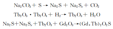
样品在烧结过程中发生如下反应[25]:
|
无水碳酸钠作为助溶剂可有效降低反应所需温度,大大节约能源。通入弱还原气氛,一方面是还原氧化铽中的四价铽,另一方面起保护作用,避免硫氧化钆在高温时氧化[26]。
2.7 Gd2O2S:Tb3+发光性能分析图 8a为Tb3+掺杂量为7%,在900 ℃下反应4 h样品的荧光激发和发射谱图。在254 nm紫外光激发下,在λ=543 nm处有对应于Tb3+(5D4→7F5)跃迁的强发射峰。在λ=412 nm、434 nm处有弱的发射峰,分别对应5D3→7F5、5D3→7F4跃迁。在λ=586 nm、622 nm处有弱的发射峰,分别对应5D4→7F4、5D4→7F3跃迁。

|
图 8 Tb3+掺杂量为7%在900 ℃下反应4 h样品的激发光谱和发射光图(a)及Tb3+能级示意图(b) Fig.8 Excitation and emission spectra (a) of the samples synthesized at 900 ℃ for 4 h, which were doped with 7% of Tb3+ and energy level diagram (b) of Tb3+ ion in the sample synthesized under the conditions as above |
能级跃迁如图 8b所示。其荧光发射过程推测为:在254 nm光激发下,Gd3+吸收能量,然后将能量以非辐射的形式传递给Tb3+的5D3能级。5D3能级的活跃电子以3种方式跃迁到低能级:
(1) 以辐射的形式从5D3能级跃迁到7Fj(j=5、4、3) 能级。
(2) 发生在5D3→5D4和7F6→7F1能级间以及5D3→7F0, 1和5D4→7F6能级间的交叉弛豫现象,Tb3+的交叉弛豫现象出现在同一Tb3+的5D3→ 5D4和7F6→7F1能级之间,正是这种交叉弛豫作用导致Tb3+的5D3能级中活跃电子数减少,5D4能级中的活跃电子数增加,发生5D4→7F j能级的发射。
(3)5D3→5D4能级间属于非辐射的跃迁方式,而5D4能级以辐射的方式跃迁到7Fj(j=6、5、4、3) 能级[27]。
3 结论以三氧化二钆、七氧化四铽和升华硫为原料,无水碳酸钠为助溶剂,采用高温固相法合成了Gd2O2S:Tb3+微米亚微米材料。反应温度、反应时间以及Tb3+掺杂量的对比实验表明,上述因素均会影响产物的荧光发光性能。通过XRD、SEM、TEM、HRTEM和荧光光谱等分析方法,对产物的物相和荧光性能进行表征,结果表明,Tb3+掺杂量为7%,在900 ℃下反应4 h的硫氧化钆样品的荧光强度最好,最强发射峰位于543 nm处,对应于Tb3+(5D4→7F5)跃迁,说明样品中Tb3+占据了Gd2O2S中部分Gd3+的晶格位点。
| [1] | Chen X, Zhao J, Yu L, Rong C, Li C, Lian S. A white light emitting phosphor Sr1.5Ca0.5SiO4:Eu3+, Tb3+, Eu2+ for LED-based near-UV chip:preparation, characterization and luminescent mechanism[J]. Journal of Luminescence, 2011, 131(12): 2697–2702. DOI:10.1016/j.jlumin.2011.06.056 |
| [2] |
吴慧钊, 张泽坤, 吴文娟. 无线平板探测器在四肢小关节X线摄影中的临床应用[J]. 中国医学影像学杂志, 2013, 21(6): 411–413.
Wu H Z, Zhang Z K, Wu W J. Clinical application of wireless flat-panel detector in X-ray photography in the small joints of the limbs[J]. Chinese Journal of Medical Imaging, 2013, 21(6): 411–413. |
| [3] | David S, Michail C, Seferis I, Valais I, Fountos G, Liaparinos P. Evaluation of Gd2O2S:Pr granular phosphor properties for X-ray mammography imaging[J]. Journal of Luminescence, 2015, 169: 706–710. |
| [4] | Fischer S, Martín-Rodríguez R, Fröhlich B, Frohlich K W, Meijerink A, Goldschmidt J C. Up conversion quantum yield of Er3+-doped β-NaYF4, and Gd2O2S:the effects of host lattice, Er3+, doping and excitation spectrum bandwidth[J]. Journal of Luminescence, 2014, 153(153): 281–287. |
| [5] |
罗昔贤. Ln2O2S:Yb, Pr(Ln=Y、La)纳米材料的上转换发光研究[J]. 功能材料, 2009, 40(6): 885–887.
Luo X X. Investigation on the up conversion properties of Ln2O2S:Yb, Pr (Ln=Y, La) nanocrystals[J]. Journal of Functional Materials, 2009, 40(6): 885–887. |
| [6] | Orlovskii Y V, Basiev T T, Pukhov K K, Polyachenkova M V, Fedorov P P, Alimov K O. Oxysulfide optical ceramics doped by Nd3+, for one micron lasing[J]. Journal of Luminescence, 2007, 125(1): 201–215. |
| [7] |
马媛媛, 何久洋, 阿孜古丽·热合曼, 巴哈德尔·肉孜, 艾尔肯·斯地克. Na4Ca4Al6Si9O24:Ce3+, Tb3+荧光粉合成及其能量传递研究(英文)[J]. 光谱学与光谱分析, 2015, 35(11): 3241–3246.
Ma Y Y, He J Y, Reheman A, Rouzi B, Sidike A. Photoluminescence of sinthetic scapolite Na4Ca4Al6Si9O24 phosphors activated with Ce3+ and Tb3+ and energy transfer from Ce3+ to Tb3+[J]. Spectroscopy and Spectral Analysis, 2015, 35(11): 3241–3246. |
| [8] | Xiong F B, Han C Y, Lin H F, Wang Y P, Lin H P, Shen H X. White light emission from novel host-sensitized single-phase Y2WO6:Ln3+, (Ln3+=Eu3+, Dy3+) phosphors[J]. Ceramics International, 2016, 42(12): 13841–13848. DOI:10.1016/j.ceramint.2016.05.189 |
| [9] | Raja A, Annadurai G, Daniel D J, Ramasamy P. Synthesis, optical and thermal properties of novel Tb3+ doped RbCaF3 fluoroperovskite phosphors[J]. Cheminform, 2016, 47(36): 654–660. |
| [10] | Osseni S A. New nanoplatform based on Gd2O2S:Eu3+ core:synthesis, characterization and use for in vitro bio-labelling[J]. Journal of Materials Chemistry, 2011, 21(45): 18365–18372. DOI:10.1039/c1jm13542b |
| [11] | Liang C J, Siao H Y. ChemInform abstract:calcining temperatures of Sr1-3xEuxDy2xAl2O4, (x=0-0.12) phosphors prepared using the potassium carbonate coprecipitation method[J]. Cheminform, 2016, 47(19): 38–45. |
| [12] | Xia T, Cao W H, Luo X X, Tian Y. Combustion synthesis and spectra characteristic of Gd2O2S:Tb3+ and La2O2S:Eu3+ X-ray phosphors[J]. Journal of Materials Research, 2005, 20(9): 2274–2278. DOI:10.1557/jmr.2005.0301 |
| [13] | Bang J, Abboudi M, Abrams B, Holloway P H. Combustion synthesis of Eu-, Tb-and Tm-doped Ln2O2S (Ln=Y, La, Gd) phosphors[J]. Journal of Luminescence, 2004, 106(3-4): 177–185. DOI:10.1016/j.jlumin.2003.09.005 |
| [14] | Zhai Y Q, Liu Y H, Meng Y, Zhang S Y. Synthesis of the red long afterglow phosphor Gd2O2S:Eu, Mg, Ti by `microwave radiation method and its luminescent properties[J]. Spectroscopy and Spectral Analysis, 2007, 27(4): 634–638. |
| [15] | Yang R Y, Lai H L. Microstructure and luminescence properties of LiBaPO4:Dy3+, phosphors with various Dy3+, concentrations prepared by microwave assisted sintering[J]. Journal of Luminescence, 2014, 145(1): 49–54. |
| [16] | Belkhiria F, Rhouma F I H, Hcini S, Daoudi M, Gammoudi H, Amlouk M. Polycrystalline La0.8Sr0.2GaO3, perovskite synthesized by sol-gel process along with temperature dependent photoluminescence[J]. Journal of Luminescence, 181: 1–7. |
| [17] | Hirai T, Orikoshi T. Preparation of Gd2O3:Yb, Er and Gd2O2S:Yb, Er infrared-to-visible conversion phosphor ultrafine particles using an emulsion liquid membrane system[J]. Journal of Colloid and Interface Science, 2004, 269(1): 103–8. DOI:10.1016/j.jcis.2003.08.026 |
| [18] | Sun W K, Hasegawa T, Abe T, Nakagawa H, Hasegawa S, Seki K. Abnormal improvement in emission of lanthanum oxysulfide phosphor La2O2S:Tb3+ synthesized by a novel method, thermal decomposition in eutectic molten salt[J]. Ceramics International, 2016, 42(8): 10389–10392. DOI:10.1016/j.ceramint.2016.03.176 |
| [19] |
梁庆华, 史瑶, 马望京, 杨新民, 李智. 混合溶剂热合成BaFBr:Eu2+微纳米晶及其性能表征[J]. 影像科学与光化学, 2012, 30(1): 33–42.
Liang Q H, Shi Y, Ma W J, Yang X M, Li Z. Mixed solvothermal synthesis and characterization of BaFBr:Eu2+ nano/micro-crystals[J]. Imaging Science and Photochemi-stry, 2012, 30(1): 33–42. DOI:10.7517/j.issn.1674-0475.2012.01.33 |
| [20] | Zhang J, Jia J. Morphologies and up-conversion luminescence of Gd4O3F6:RE3+ (RE=Yb, Er, Ho and Tm) phosphors by Hydrothermal Synthesis[J]. Journal of Luminescence, 2016, 174: 1–5. DOI:10.1016/j.jlumin.2016.01.030 |
| [21] | Song Y, You H, Huang Y, et al. Highly uniform and monodisperse Gd2O2S:Ln3+ (Ln=Eu, Tb) submicrospheres:solvothermal synthesis and luminescence properties[J]. Inorganic Chemistry, 2010, 49(24): 11499–504. DOI:10.1021/ic101608b |
| [22] |
范艳玲, 吕雪莉, 刘艺芳, 孟凡成, 时孟琪, 张香兰. 微乳液法制备纳米二氧化硅研究进展[J]. 中国科技博览, 2010(25): 16–18.
Fan Y L, Lv X L, Liu Y F, Meng F C, Shi M Q, Zhang X L. Study on synthesis technology for nano-sized SiO2 powder by microemulsion method[J]. China Science and Technology Expo, 2010(25): 16–18. |
| [23] |
陈万平, 周阿红. X射线荧光粉的发光与应用[J]. 怀化学院学报, 2015(5): 27–32.
Chen W P, Zhou A H. Luminescence mechanism and application in medical imaging of X-ray phosphors[J]. Journal of Huaihua University, 2015(5): 27–32. |
| [24] |
郑梁, 张芳芳, 宋开新, 郑鹏, 吴松, 郑海杰. Li2Sr0.995-xSiO4:0.005Eu2+, xLa3+荧光粉的晶体结构与发光特性[J]. 中国稀土学报, 2013, 31(5): 30–36.
Zheng L, Zhang F F, Song K X, Zheng P, Wu S, Zheng H J. Luminescence characteristics and crystal structures of phosphors Li2Sr0.995-xSiO4:0.005Eu2+, xLa3+[J]. Journal of the Chinese Society of Rare Earths, 2013, 31(5): 30–36. |
| [25] |
刘应亮, 宋春燕, 张静娴, 袁定胜, 黄浪欢, 容建华, 张俊文. Er3+, Ho3+和Tm3+在硫氧化钆中的余辉发光[J]. 无机化学学报, 2005, 21(6): 905–909.
Liu Y L, Song C Y, Zhang J X, Yuan D S, Huang L H, Rong J H, Zhang J W. After glow emission of Er3+, Ho3+ and Tm3+ in gadolinium oxysulfide[J]. Chinese Journal of Inorganic Chemistry, 2005, 21(6): 905–909. |
| [26] | Haynes J W, Brown J J, Haynes J W, Brown J J. Preparation and luminescence of selected Eu3+ activated rare earth-oxygen-sulfur compounds[J]. Journal of the Electrochemical Society, 1968, 115(10): 1060–1066. DOI:10.1149/1.2410877 |
| [27] |
郭楠, 郭坤, 盛野, 邹海峰. Tb3+和Gd3+掺杂的纳米TiO2薄膜的制备及发光性能与发光机理[J]. 吉林大学学报理学版, 2009, 47(2): 367–375.
Guo N, Guo K, Sheng Y, Zou H F. Optical properties and energy transfer mechanism of Tb3+/Gd3+ co-doped nanometer TiO2 thin film[J]. Journal of Jilin University, 2009, 47(2): 367–375. |